
HI guys is Zadi, welcome to another post, today I will be talking about Normal first-trimester ultrasound and all you need to know, so stay with me !!!!
OK, so assuming that you have a positive pregnancy test, HCG ( pregnancy hormone ) blood work that confirms pregnancy, next thing is an ultrasound to rule out gestational age, estimated due date, how many babies and also maternal organs are check to rule out Fibroids, ovarian cysts etc..
If you haven’t seen my previous posts about fibroids and ovarian cysts I will link down below for you:
All you need to know about Fibroids.
All you need to know about Ovaries, normal hormonal cycles, and common cysts.
So now back to the post, what are you going to see on the first ULTRASOUND scan ( 6 weeks of gestation )??

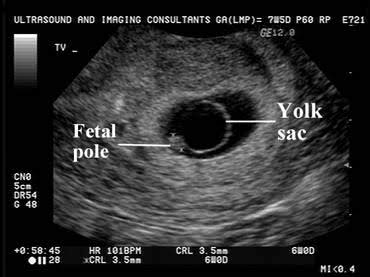
Gestational sac: The first structure seen on ultrasound, the gestational sac can be visualized as early as 5 weeks by Transvaginal technique. A normal gestational sac can be round or oval and is located within the fundus or mid portion of the cavity in the uterus.
 Gestational sac within the cavity.
 Ultrasound image of the gestational sac and yolk sac.
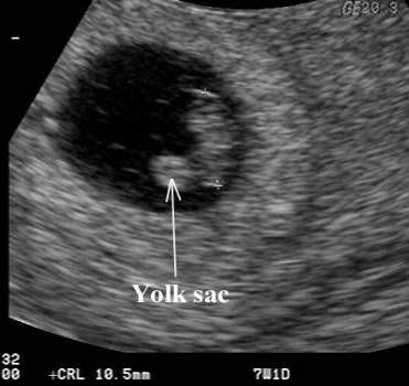
Yolk sac: The yolk sac is the first structure visible within the gestational sac. The yolk sac should always be seen when the gestational sac measures greater than 1.0 cm or 10 mm.
A normal yolk sac is round and should measure less than 6 mm. If the yolk sac measures more than 6 mm, is bizarre in shape or is calcified follow up scan is required since most pregnancies with Abnormal yolk sacs will fail.
 Normal looking yolk sac.
 Enlarged yolk sac.
 Calcified yolk sac.
 Yolk sac with a bizarre shape.
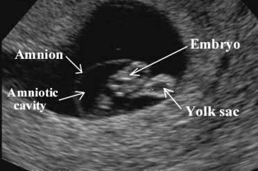
Embryo: The second structure that becomes visible within the gestational sac is the embryo. Embryonic cardiac activity should always be seen when an embryo measures greater than 5 mm.
A normal fetal heart rate usually ranges from 115 to 160 beats per minute during the first weeks of gestation, It is measurable with ultrasound from around 6 weeks, the normal range varies during gestation, increasing to around 150 to 175 beats per minute at 10 weeks and decreasing to around 130 beats per minute at term.
A heartbeat of 100 BPM or less In the first ultrasound may indicate a pregnancy that is going to fail however a follow up is always recommended.

Ultrasound image of an Embryo at about 6 weeks of gestation.
Membranes: The amnion grows against the chorion and the membranes eventually fuse, usually by 17 weeks.
Most COMMON and usually harmless cause of vaginal bleeding on the first trimester is Subchorionic bleedings, it resolves by itself however precautions have to be follow.

Ultrasound image of subchorionic bleeding.
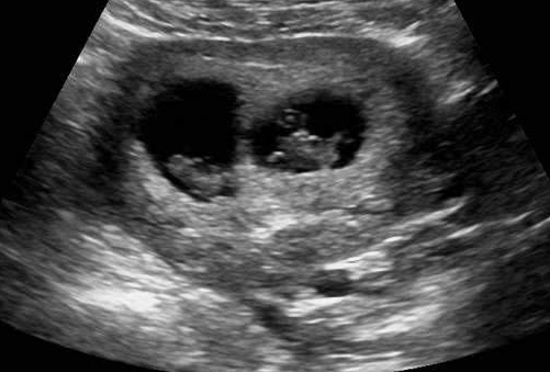
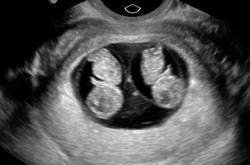
And remember if you see an image like this one:
you guess it !!!! YOU ARE HAVING TWINS…
 Ultrasound image of Fraternal twins. ( I will explain this in another post )
 Ultrasound image of Identical twins.

So much information, very interesting
LikeLiked by 5 people
Thank you my friend, is a compliment coming from someone like you, I really enjoy your blog posts.
LikeLiked by 2 people
You are welcome
LikeLiked by 4 people