
Hey guys, welcome to another post, today I want to briefly talk about the Abnormal first trimester ultrasound before I continue to weekly ultrasounds, as usual, I will be including ultrasound pictures on my post, in case you haven’t seen my previous article about Normal first trimester ultrasound I will link it down below:
Normal first trimester ultrasound, scan at 6 weeks of gestation.
First trimester ultrasound measurements, dating, and guidelines…
Pregnancy failure is a common problem in the first trimester with failure rates approaching 25%. A threatened abortion is defined as bleeding and cramping in the first 20 weeks of pregnancy. Ultrasound plays a key role in evaluating women with threatened abortion since HCG levels do not always correlate with a specific diagnosis.
Subchorionic hematoma or Subchorionic bleedings.

Is the most common sonographic abnormality in the presence of a live embryo. Vaginal bleeding affects 25% of all women during the first trimester. In women whose sonogram shows a Subchorionic hematoma, the outcome of the fetus depends on the size of the hematoma, the mother’s age and the fetus gestational age.
If the Subchorionic hematoma appears in the late first or second trimester the risks for miscarriage, stillbirth, placental abruption or preterm labor are increased.
However small asymptomatic Subchorionic hematoma vs Subchorionic bleeding associated with No other complications can resolve by itself.
Anembryonic pregnancy ( Blighted ovum).

Is a form of failed pregnancy defined as a gestational sac in which the embryo failed to develop. A large gestational sac without the visualized embryo is unequivocal evidence of a failed anembryonic pregnancy.
Embryonic demise and Bradycardia.

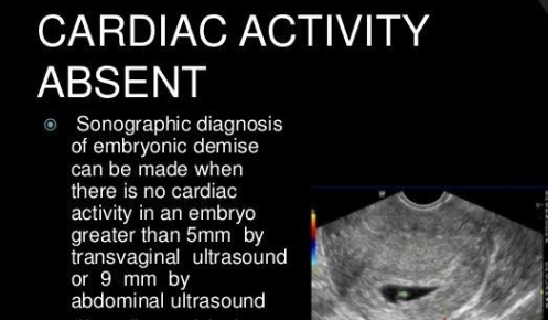
The most convincing evidence that pregnancy has failed is the documentation of embryonic demise. As stated previously, all embryos greater than 5 mm in size should demonstrate cardiac activity. Embryonic bradycardia is a poor prognosticator of pregnancy viability and needs follow up. An embryonic heart rate less than 90 beats per minute, in embryos less than 8 weeks is associated with 80% rate of eventual embryonic demise.
Ectopic pregnancy.
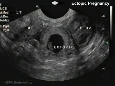
Tubal ectopic pregnancy located at the LT adnexa.
The most reassuring sign that an ectopic pregnancy is not present is the sonographic demonstration of a normal intrauterine pregnancy.
Which means that when the HCG levels are 1500 IU or more an intrauterine pregnancy has to be seen, if not the possibility of an Ectopic pregnancy is really high, Follow up with HCG in blood and Ultrasound are highly recommended.
Patients that are in high risk for ectopic pregnancy have:
*History of pelvic inflammatory disease ( PID )
*Previous ectopic pregnancy.
*Infertility.
*Tubal surgery.
Transvaginal ultrasound has an accuracy of 90% and should be routinely be used in the evaluation for ectopic pregnancy.
A variety of uterine findings can be seen with ectopic pregnancies. The may be empty or contain endometrial fluid collection, this should not be confused with an intrauterine gestational sac.
The most common adnexal finding is a complex mass which represents hemorrhage. Other adnexal findings included a normal adnexa or a well formed adnexal ring with or without a yolk sac or embryo. The posterior uterine pouch or pouch of Douglas should be carefully investigated since complex peritoneal fluid may be the only finding in 15% of ectopic pregnancies.

OK so I am ending my post here, on the next one I will be talking probably about Twin pregnancies, and the following week I will be going week by week on scanning, if you stay until the end, thanks and I hope you come back…
feel free to comment if you have any questions.
xo zadi.

lots of important information here
LikeLiked by 7 people
Thanks my dear
LikeLiked by 2 people
I had the asymptomatic type of bleeding… luckily she told me that so I didn’t panic
LikeLiked by 4 people
You see, I am so glad to know. As an ultrasound tech I know that sometimes is harmless, however I see how all the patients get to the ultrasound freaking out. That’s why I wanna to do a post like this one to give peace of mind.
LikeLiked by 2 people
Very important!
LikeLiked by 3 people